30. Review on: SEIRA spectroscopy

SEIRA spectroscopy is an important tool in the molecular analysis of surfaces. We review the basis for SEIRA, common approaches as well as important case studies in the fields of electrochemistry, trace analysis and (bio)functional investigations.
Kozuch et al., Nat. Rev. Methods Primers, 2023
Surface-enhanced infrared absorption spectroscopy.
DOI: 10.1038/s43586-023-00253-8
15. Expanding the voltage Window Using Diazonium-Derived Interfaces for Studies of Redox Proteins

The application of redox proteins interfaced on sulfur-based self-assembled monolayers (SAM) is severely restricted the SAM stability. Using diazonium-derived SAMs forming a covalent Au-C bond the voltage window is increased by up to 1 V. We demonstrate this approach using an oxygen-tolerant hydrogenase.
Harris et al., ACS Appl. Mat. Interfaces, 2018
In situ spectroelectrochemical studies into the formation and stability of robust diazoniumderived interfaces on gold electrodes for the immobilization of an oxygen-tolerant hydrogenase.
DOI: 10.1021/acsami.8b02273
13. Monitoring the Hydrogen evolution by Bio-inspired cobalt hangman porphyrins under operating conditions
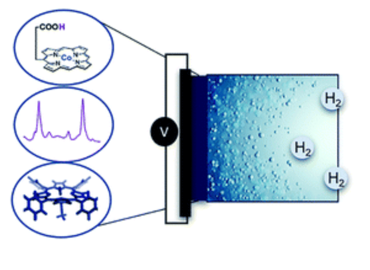
Cobalt hangman complexes are promising bio-inspired catalysts for dihydrogen production, yet their electrocatalytic performance is still a topic of dispute. Using surface-enhanced resonance Raman (SERR) spectro-electrochemistry we provide insight into the reaction mechanism at electrodes under turnover conditions.
Kielb et al., Catal. Sci. Technol., 2018
Hydrogen evolution by cobalt hangman porphyrins under operating conditions studied by vibrational spectro-electrochemistry.
DOI: 10.1039/C7CY02253K
8. How does the 2nd coordination sphere in biomimetic complexes control electron transfer?
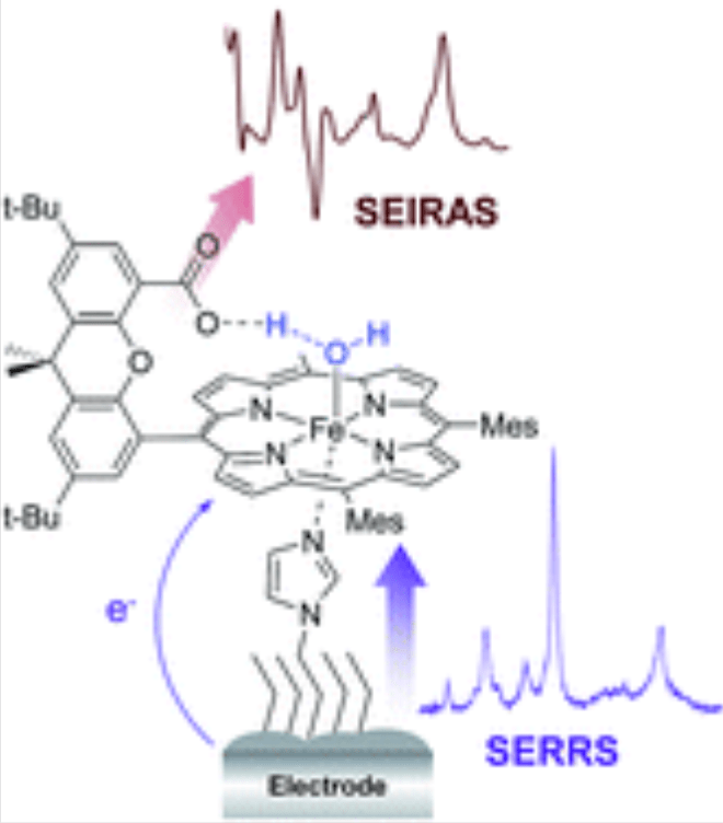
Iron hangman complexes exhibit improved catalytic properties for O2 and H2O2 reduction, which are attributed to the presence of a proton donating group in the 2nd coordination sphere of the catalytic metal center. We propose a PCET that is modulated by the protonation state of the acid hanging group via hydrogen bond interactions.
Ly et al., Chem. Sci., 2015
2nd coordination sphere controlled electron transfer of iron hangman complexes on electrodes probed by surface-enhanced vibrational spectroscopy.
DOI: 10.1039/C5SC02560E
7. A simple Method to create Ag Polyhedrons on Gold as a Platform for Surface-enhanced spectroscopies

We create polyhedron Ag nanostructures on top of Au electrodes via step-wise electrodeposition and analyze this system as substrates for surface-enhanced Raman spectroscopy.
Kozuch et al., Phys. Chem. Chem. Phys., 2015
Calculating average surface-enhancement factors of randomly nanostructured electrodes by a combination of SERS and impedance Spectroscopy.
DOI: 10.1039/C4CP05015K
5. Using TiO2 electrodes as substrates for Surface-enhanced Raman Spectro-electrochemistry of immobilized Redox proteins

Nanostructured silver and gold substrates are the standard material to obtain surface-enhancement for Raman spectroscopy of immobilized proteins. Here, we use nanostructured titanium dioxide (TiO2) electrodes to probe the electron-transfer process of cytochrome b5 (cyt b5) by surface-enhanced resonance Raman (SERR) spectroscopy.
Han et al., Small, 2013
Potential-Dependent surface-enhanced resonance Raman spectroscopy at nanostructured TiO2: a case study on cytochrome b5.
DOI: 10.1039/C4CP05015K
2. Biocompatible Surface-enhanced resonance Raman Spectroscopy using Silica-coated Ag nanoparticles

We synthesized silica-coated Ag nanoparticles with defined surface plasmon resonances to selectively detect and analyze protein cofactors via surface enhanced resonance Raman spectroscopy. The silica coating does not diminish the optical amplification considerably, but minimizes unwanted interactions between the protein and the nanoparticle.
Sivanesan et al., RSC Adv., 2012
Tailored silica coated Ag nanoparticles for non-invasive surface enhanced Raman spectroscopy of biomolecular targets.
DOI: 10.1039/C1RA00781E
1. Tuning the optical Properties of Ag Nanoparticles for surface-Enhanced Resonance Raman Spectroscopy of Proteins

We present a preparation procedure for small sized biocompatibly coated Ag nanoparticles with tunable surface plasmon resonances. The conditions were optimised with respect to the resonance Raman signal enhancement of heme proteins and to the preservation of the native protein structure.
Sivanesan et al., Chem. Commun., 2011
Functionalized Ag nanoparticles with tunable optical properties for selective protein analysis.
DOI: 10.1039/C0CC05058J
